Home
Capital Campaign
Who we are

The Foundation for Veterinary Dentistry was established in 2005 to provide a vehicle for charitable donations that fund research and education in the field of veterinary dentistry. The Foundations scope and involvement in veterinary dentistry has grown and evolved over the years. It now manages both the Journal of Veterinary Dentistry and the annual Veterinary Dental Forum, as well as supports numerous other philanthropic endeavors.
That is why in 2019 the Foundation established a long-term endowment fund through the Community Foundation of Northern Colorado. Each year, a percentage of the average fund balance will be available for distribution to support our Foundation Mission; to support veterinary dental research and education. This endowment will provide essential stability, facilitate the strategic use of funds and allow our organization to plan on the income from one year to the next. This endowment fund also presents an excellent option for donors who want to make legacy or planned gifts that will keep providing funds far into the future.
The goal is to establish a fund of $1 million dollars in an endowed (protected) fund to ensure future financial support for research and education in the field of veterinary dentistry. This would provide ongoing funding of $30,000 to $50,000 each year.
Sponsoring dental education throughout America – see more

Funding dental education via Regional Collaborative Dental Education Centers – see more

Funding Grants to help provide better healthcare for animals
Cone-beam computed tomography and pathology of canine
temporomandibular joint osteoarthritis: an agreement study
Principal Investigator: Boaz Arzi DVM, DAVDC, DEVDC, Founding Fellow AVDC OMFS, School of Veterinary Medicine, University of California, Davis
Introduction
Research by our group indicates that TMJ disorders are common in dogs. Specifically, 81% of dogs undergoing computed tomography (CT) imaging of the skull were found to have findings consistent with TMJ osteoarthritis (OA). The routine use of CT and cone beam CT (CBCT) imaging for the diagnosis of TMJ disorders in dogs is gaining rapid acceptance but the sensitivity of CBCT for detecting pathology in the TMJ of dogs has not been studied.
This study sought to identify CBCT imaging findings that correlate with gross and microscopic evidence of TMJ OA, and to better characterize the role of the TMJ disc in canine TMJ OA. The proposed research is the first systematic comparison of CBCT and pathologic evidence of TMJ OA. Correlating CBCT findings with TMJ pathology will aid interpretation of the likely severity of TMJ OA in individual dogs. Additionally, we expect that some specific CT findings are likely to be associated with more severe joint pathology while other findings are more likely to be incidental. Understanding the severity of TMJ pathology based on CT findings will improve diagnosis of TMJ OA and help clinicians identify dogs with potentially clinically important disease. In addition, understanding TMJ disc degeneration associated with OA may lead to recommendations for additional diagnostic tests (e.g., TMJ arthrography or MRI) in clinical patients when TMJ disorders are suspected.
The combined findings of this research will expand our general understanding of canine TMJ OA, improve its diagnosis in clinical patients, and lead to future research toward treating dogs with clinically significant TMJ OA.

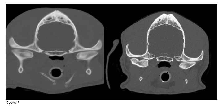
Figure 1: Cone Beam Computed Tomography section of the healthy TMJ of a dog (right image).
On the left side, there is severe focal arthritic lesion of the left TMJ
Identification of Targetable Activating Mutations in
Canine Acanthomatous Ameloblastoma
Dr. Santiago Peralta, DVM, Dipl. AVDC, FF-AVDC-OMFS, and his collaborators received a $10,000 grant from the Foundation for Veterinary Dentistry
The project was immensely successful and led to important advances and discoveries regarding the molecular mechanisms involved with this common and locally destructive canine oral tumor. Importantly, they discovered that an acquired mutation in the HRAS gene (i.e. p.Q61R) is present in the vast majority of tumors. This discovery is of major veterinary and translational significance because it will serve as the basis for the development of novel precision-medicine based therapies in dogs in the near future. Moreover, such therapies …..
Thank you to our
2025 Titanium Member Contributors
Dr. Josephine Banyard
Dr. Jan Bellows
Dr. Ian Bishop
Dr. Stanley Blazejewski
Dr. Glenn Brigden
Dr. Cindy Charlier
Companion Animal Dentistry of Kansas City
Dr. William Gengler
Dr. Paul Hobson
Dr. Stephen Juriga
Dr. Kenneth Lee
Dr. Milinda Lommer
Dr. Sue McTaggart
Dr. Katherine Queck
Dr. Erich Rackwitz
Dr. Billy Scott
Dr. Bonnie Shope
