Cone-beam computed tomography and pathology of canine temporomandibular joint osteoarthritis: an agreement study
Principal Investigator: Boaz Arzi DVM, DAVDC, DEVDC, Founding Fellow AVDC OMFS, School of Veterinary Medicine, University of California, Davis
Introduction
Research by our group indicates that TMJ disorders are common in dogs. Specifically, 81% of dogs undergoing computed tomography (CT) imaging of the skull were found to have findings consistent with TMJ osteoarthritis (OA). The routine use of CT and cone beam CT (CBCT) imaging for the diagnosis of TMJ disorders in dogs is gaining rapid acceptance but the sensitivity of CBCT for detecting pathology in the TMJ of dogs has not been studied.
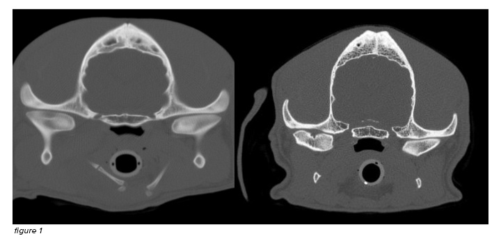
This study sought to identify CBCT imaging findings that correlate with gross and microscopic evidence of TMJ OA, and to better characterize the role of the TMJ disc in canine TMJ OA. The proposed research is the first systematic comparison of CBCT and pathologic evidence of TMJ OA. Correlating CBCT findings with TMJ pathology will aid interpretation of the likely severity of TMJ OA in individual dogs. Additionally, we expect that some specific CT findings are likely to be associated with more severe joint pathology while other findings are more likely to be incidental. Understanding the severity of TMJ pathology based on CT findings will improve diagnosis of TMJ OA and help clinicians identify dogs with potentially clinically important disease. In addition, understanding TMJ disc degeneration associated with OA may lead to recommendations for additional diagnostic tests (e.g., TMJ arthrography or MRI) in clinical patients when TMJ disorders are suspected.
The combined findings of this research will expand our general understanding of canine TMJ OA, improve its diagnosis in clinical patients, and lead to future research toward treating dogs with clinically significant TMJ OA.

Figure 1: Cone Beam Computed Tomography section of the healthy TMJ of a dog (right image). On the left side, there is severe focal arthritic lesion of the left TMJ

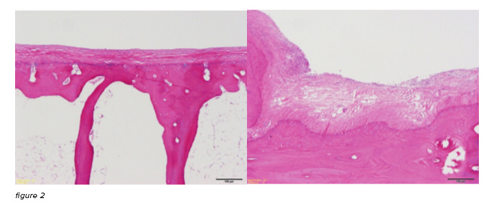
Figure 2: Histological images of the TMJ of 2 dogs. On the right, normal articular surface of the mandibular head on the condylar process of a dog. On the left, moderate to severe articular surface lesion with disruption of the articular fibrocartilage.

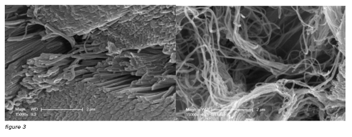
Figure 3: Scanning Electron Microscopy of the TMJ disc from 2 dogs. On the left there I normal fiber contour and arrangement. On the right, there is moderate-severe disruption of collagen fibers as a result of degenerative changes of the TMJ.
